
Acute pulmonary edema, commonly abbreviated as APE, refers to a sudden accumulation of fluid in the pulmonary alveoli. This phenomenon prevents normal gas exchange and causes acute respiratory failure. Understanding the types of APE in medicine allows for rapid management, as treatment varies significantly depending on the underlying mechanism.
Alveolo-capillary mechanism: what happens in the lung during APE
In a healthy lung, a thin membrane separates the air in the alveoli from the blood circulating in the capillaries. This alveolo-capillary membrane allows oxygen to pass into the blood and facilitates the removal of carbon dioxide. The balance relies on a play of pressures: hydrostatic pressure pushes fluid out of the vessels, while oncotic pressure (related to plasma proteins) holds it inside.
When this balance is disrupted, fluid invades the interstitial space and then the alveoli. Alveolar flooding drastically reduces the surface area available for gas exchange. The patient then develops intense dyspnea, often worsened when lying down, accompanied by sweating, cyanosis, and sometimes pink frothy sputum.
Two main mechanisms can cause this disruption of balance, and it is precisely this distinction that underpins the classification into types of APE. To better understand the main types of APE in medicine, one must first distinguish between an elevation of pressure upstream of the capillary and a direct destruction of the membrane itself.
Cardiogenic APE: when the left heart is involved
Cardiogenic APE represents the most commonly encountered form in clinical practice. Its origin lies in a failure of the left heart, which no longer effectively ejects blood into the systemic circulation. Blood then stagnates upstream, in the pulmonary veins and then in the capillaries, causing an elevation of hydrostatic pressure.
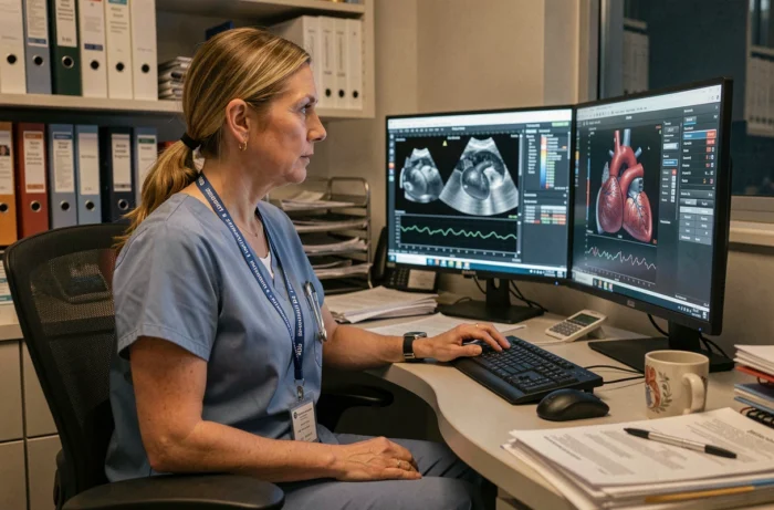
This overpressure forces plasma fluid to cross the capillary wall into the alveoli. The alveolo-capillary membrane remains intact: it is the excessive pressure that is responsible for the leakage.
Common causes of cardiogenic APE
- Decompensation of a pre-existing left heart failure, often triggered by dietary indiscretion, discontinuation of treatment, or an intercurrent infection
- Acute coronary ischemia (myocardial infarction) that suddenly weakens the contraction of the left ventricle
- A rapid cardiac rhythm disturbance or an acute valvular pathology (mitral chord rupture, endocarditis) disrupting intracardiac blood flow
- An acute volume overload, for example during a too-rapid intravenous infusion in a patient whose cardiac function is already compromised
Treatment focuses on reducing fluid overload (diuretics) and supporting cardiac function. Sitting with legs dangling immediately relieves venous return to the heart.
Lesional APE: direct damage to the pulmonary membrane
In lesional APE, the problem does not stem from the heart. The alveolo-capillary membrane is directly damaged, making it permeable to fluid and plasma proteins. Cardiac pressures remain normal, but fluid still infiltrates the alveoli due to tissue destruction.
This form corresponds to what medical literature also refers to as non-cardiogenic pulmonary edema. In its most severe presentations, it evolves into an acute respiratory distress syndrome (ARDS), characterized by severe hypoxemia refractory to simple oxygen therapy.
Origins of the lesional damage
The causes are varied and affect either the lung directly or the body systemically:
- Severe infectious pneumonia or inhalation of gastric fluid, which destroy the alveolar epithelium through direct aggression
- Extrapulmonary sepsis, where the systemic inflammatory response alters capillary permeability throughout the body, including the lungs
- Inhalation of toxic substances, drowning, polytrauma, or fat embolism, all situations where the alveolar barrier is compromised by an inflammatory or mechanical mechanism
The overall definition of ARDS published in 2024 has broadened the diagnostic criteria. It now includes criteria applicable to certain non-intubated patients, under high-flow oxygen therapy or non-invasive ventilation. It also allows the use of the SpO2/FiO2 ratio under defined conditions, facilitating diagnosis outside of intensive care units.
Mixed forms and differential diagnosis of APE
The cardiogenic/lesional distinction, as clear as it may be in theory, often blurs in clinical practice. Both mechanisms can coexist in the same patient. A heart failure patient hospitalized for decompensation may simultaneously develop a nosocomial pneumonia that damages the alveolar membrane. The clinical picture then combines overpressure and membrane destruction.
This is why the diagnosis relies on a bundle of arguments: clinical history, physical examination, biology, imaging. A chest X-ray shows bilateral alveolar opacities in both cases, making it insufficient on its own to make a definitive diagnosis.
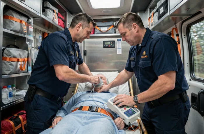
Bedside lung ultrasound has taken on an increasing role in initial assessment. B-lines (vertical artifacts visible on ultrasound) indicate interstitial congestion, but they are not specific to heart failure: they are also found in pneumonias and lesional APE. Associated cardiac ultrasound allows for the evaluation of left ventricular function and filling pressures, providing a decisive argument in favor of a cardiogenic origin.
Lung ultrasound is not limited to initial diagnosis. It is also used to monitor decongestion under treatment, quantifying the regression of B-lines over time.
The stakes of this differential diagnosis are directly therapeutic. Administering high-dose diuretics in lesional APE without a cardiac component risks worsening hypovolemia. Conversely, fluid resuscitating a patient in cardiogenic APE precipitates respiratory distress. The type of APE dictates every therapeutic decision, from the first hour until hospital discharge.